For years doctors could only listen to the sound of your heart with a stethoscope to determine it was functioning well and even today doctors are often pictured with a stethoscope around their neck. In fact, 30-40 years ago ultrasound was introduced to look at the function of the heart and today every cardiologist has a 2D ultrasound machine and uses it many times a day. Like most medical technology, it is a work in progress and innovations are needed to overcome the device’s limitations. One limitation is the analysis of the 16 different 2D slices (also called views), which are taken in a standard echocardiogram. Everyone is different and due to the physics of sound and the anatomy of people, it is often impossible to get clear images for all 16 views. It then becomes very difficult for the cardiologist to assess all aspects of heart function. This limitation has been overcome by a small Canadian Company, Ventripoint Diagnostics Ltd (TSXV:VPT).
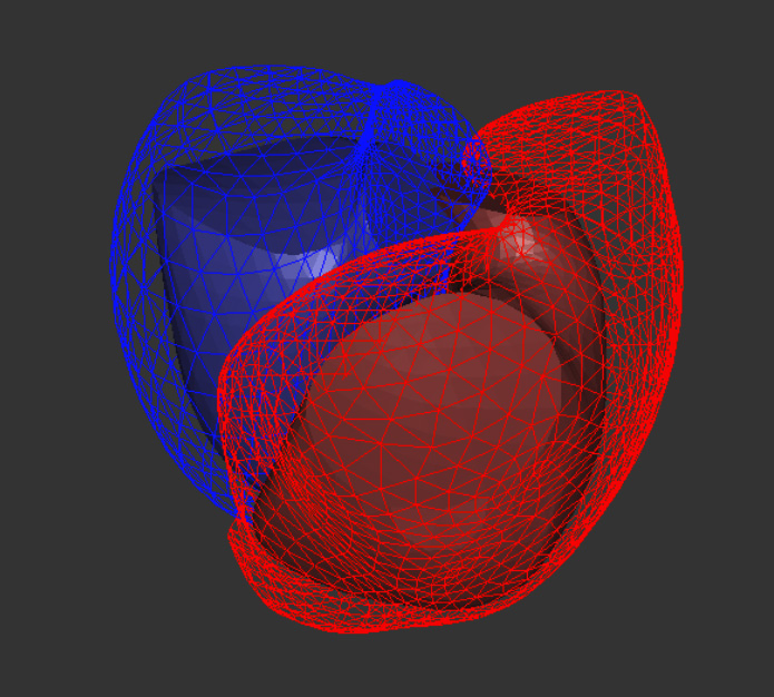
Ventripoint has been working on this problem for over 15 years and has developed and patented an artificial intelligence (AI) technology called knowledge-based reconstruction (KBR). This revolutionary approach uses a library of heart images – both normal and dysfunctional, to help analyze a patient’s heart. using the images from conventional 2D ultrasound. The Company has scoured the world for the oddest hearts as it has discovered the best way to deal with anyone’s heart is to have the extreme hearts in the library. This focus has allowed their whole heart analysis system to yield results equivalent to MRI from ultrasound images. The approach also works on MRI images and even 3D ultrasound images, but these other imaging modalities have their own set of limitations and so 99% of heart analysis is done with 2D ultrasound.
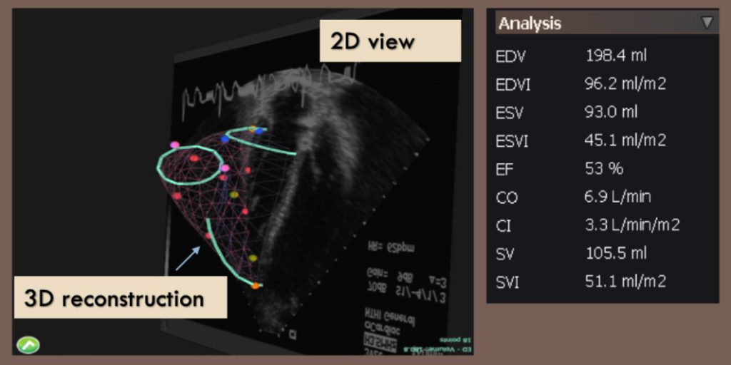
The key to Ventripoint’s AI approach is the ability to use sparse data to build a 3D model of the heart. The sparse data concept is key as simply being able to identify a few points in each view is enough for the KBR to build an accurate model. It does this extremely accurately and so the cardiologist can be confident in the results. This approach is obvious to clinicians who look at heart images every day; plus the technique is very easy to learn. The company just announced a study where 100% of clinicians were able to learn and operate the newest model, the VMS+3.0, with just three hours of training. Even better – they were able to remember how to do it a few days later without assistance.
A special focus of the company has been pediatric heart disease. Most infants born with a heart problem have defective right hearts. Historically it has been particularly challenging to get clear, readable images of the right heart. So, it is hard to analyze using ultrasound images. Placing infants and toddlers in an MRI is the only other way to determine the full nature of their heart problems and this is both dangerous and expensive. It was this puzzle that inspired Dr. George Adams, the CEO of Ventripoint, and sparked him to search for better heart analysis approach. After talking to pediatric cardiologists, who said one of the hardest parts of their jobs was to decide to do an MRI on a child. It takes 1.5 to 2.5 hours inside an MRI to capture the same 16 views you can get with ultrasound in 10 minutes. Obviously, an MRI is traumatic to a child as they need to be sedated and put on a mechanical ventilator and forced to hold their breath during image acquisition. You can see why the decision is so stressful for doctors and parents.
Here is what Dr. Luc Mertens, Head, Echocardiography Cardiology at The Hospital for Sick Children has to say about the VMS:
“We found an excellent agreement between both techniques and, based on these findings, decided to introduce the method in our Echocardiography Laboratory. The first results of a clinical trial to evaluate feasibility, reproducibility and accuracy of the system are really impressive and demonstrate that the VMS is a robust clinical tool. I see a multitude of potential applications for the VMS that can significantly impact the care of patients with heart disease.”
A Picture of the Whole Heart
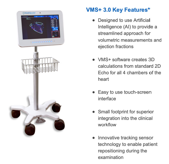
So where is Ventripoint in its quest to change the way hearts are analyzed? Having shown in many clinical studies and with the approval of Health Canada, the US-FDA and the European regulatory bodies, the VMS yielded accurate and equivalent results to the gold standard MRI for one chamber of the heart, the right ventricle, they embarked upon expanding the system to image all 4 chambers of the heart – the whole heart. This expansion allowed the VMS system to analyze all types of heart dysfunctions in adults and children, and not just congenital heart disease in children. This milestone was achieved last year and testing of the system on adults with heart problems shows the same excellent results equivalent to MRI. The Company has now developed a smaller, more user-friendly VMS+3.0 and has submitted for approval to sell this new model. Consequently, hospitals, clinics and cardiologist offices have been waiting for this next-generation device. The company is at a tipping point with a validated technology that addresses a large unmet medical need with a global market.
Time to Sound off to the World about a Better Way
The largest heart ultrasound congress in the world is happening in Portland Oregon, right now, and Ventripoint is exhibiting the new VMS+3.0 to the 17,000 attendees. They have live scanning in the booth, so anyone can try it out. There will also be a scientific presentation by the cardiology group from Mazankowski Heart Institute in Edmonton to report on their study using the VMS+ to analyze “unreadable” echocardiograms. The hope is the cardiologist will be able to find enough recognizable points in the views to properly analyze the function of the left ventricle in technically challenging patients, who yielded such blurry images that they were deemed “unreadable”. This happens often with up to 25% of patients in some conditions. One approach to obtain better images is to inject contrast media into patients while redoing the ultrasound scanning and quickly retake just the two best views. This takes extra time, is uncomfortable for patients and is dangerous as some people are allergic to the contrast dye. It also adds extra cost and the requirement for a nurse to do the injection. All of this could be eliminated by simply analyzing the original views using the VMS+3.0. We await the report on the study at the conference.
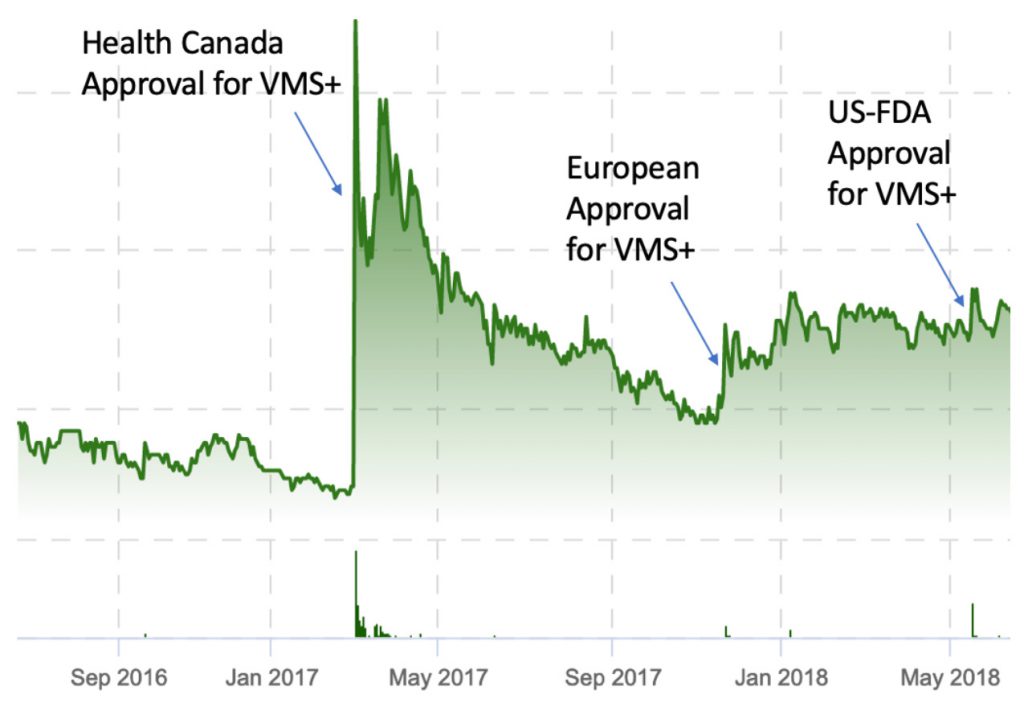
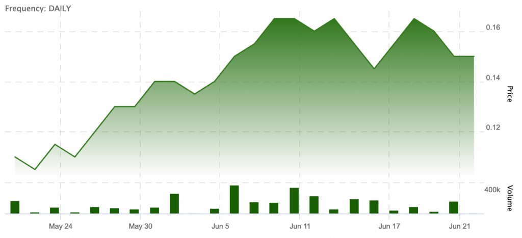
The Company has applied for regulatory approval in Canada, United States and Europe and expects to receive the approvals in the next few weeks. The stock price has a jump on announcement of regulatory approvals. This happened every time the company has introduced its next generation of products with increases up to 9 times the price prior to the announcement with exceptional liquidity.
The stock price has started to move up in the last few weeks in anticipation and now would be the ideal time to invest in Ventripoint (TSXV:VPT, OTCQX:VPTDF).
Sounds Like an opportunity!